


Facilities & Equipments
New Revolution in Retinal OCT (OPTICAL COHERENCE TOMOGRAPHY) Angiography
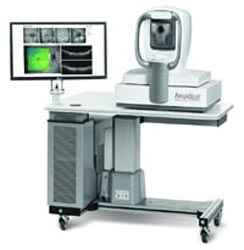
This equipment is used to visualize each of the retinas distinctive layers. We can perform retina scanning, diagnose macula disease, and diagnose diseases in optical disc. It Helps to diagnose Glaucoma, intervention in Diabetic Retinopathy, early signs of Age Related Retina Macular Degeneration and Keratoconus using Epithelial thickness mapping.
Advanced Features
- 3D, Highest Resolution Imaging of smaller blood vessels in full details.
- Retinal Angiography without dye injection.
- No Dilation drips required.
- Quick and easy imaging.
- Non Invasive.
FFA (FUNDUS FLUORESCEIN ANGIOGRAPHY)

It is a technique for examining the circulation of the retina and choroid (parts of the fundus) using a fluorescence dye and a specialized camera. Sodium fluorescein is added into the systemic circulation, the retina is illuminated with blue light at a wavelength of 490 nanometers, and an angiogram is obtained by photographing the fluorescent green light that is emitted by the dye. It identifies leakage in blood vessels. It is use to diagnose prolifrative and non-proliferatve diabetic retinopathy
Advanced Features
- Colour, red-free, fluorescein standard
- ICG, autoflourescence optional
- 20, 35, 50 angles of coverage
- Easy to use touch screen control panel
- Easy focusing system with split focus bars
- Can be used with a wide range of digital cameras
- IMAGEnet i-base connection
HRA FFA (HEIDELBERG RETINAL ANGOGRAPHY)
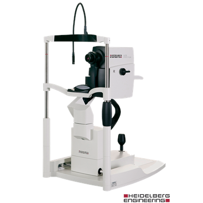 This equipment is used to visualize retinal blood vessels with the help of fluorescent. We do not have to inject the dye to the patient. But, to identify the exact point of leakage Indocyanin green dye is injected. The dye makes the blood vessels white and the blood appears black in color. Thus, the doctor is able to identify the leakage is from which vessel.
Product Features
- TruTrack™ active eye tracking: actively follows the patient’s eye during the scan, minimizing motion artifact.
- It combines multiple scans taken at the same location and eliminates noise from the image.
- Confocal Scanning Laser Ophthalmoscope: for high resolution fundus imaging.
- BluePeak™ with RegionFinder™: to monitor RPE health in conditions such as AMD and hydroxychloroquine administration.
- Five (5) imaging modes: infrared (IR), BluePeak™ blue laser autofluorescence, blue reflectance (BR) (this mode is sometimes referred to as red-free), fluorescein angiography (FA) and ICG angiography (ICGA).
- Movie image capture: for high speed video angiography.
- Optional non-contact ultra-widefield angiography: offers evenly illuminated, undistorted, high contrast images in the far periphery.
- Fundus image field of view: from 15° up to 150° with optional lenses.
- Network ready: remote image review and optional EHR/PACS interface.
- Upgradable: the following additional imaging modes can be added to this system: Spectral Domain OCT.
INTRALASE FEMTOSECOND LASER
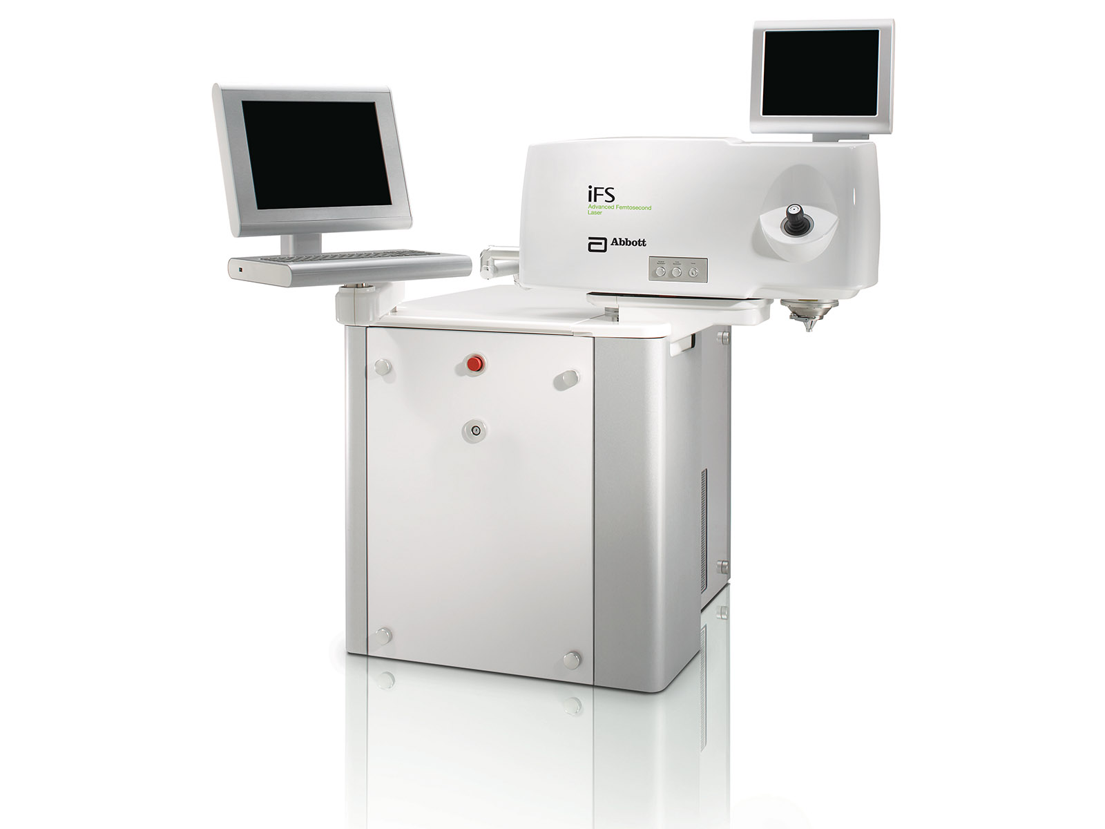
It is a comprehensive platform built on IntraLase Technology legacy of surgical precision. Delivering fully individualized procedures from LASIK flaps to inlay pocket creation, the iFSLaser is the ideal choice for every ophthalmic subspecialty.
Advantages of femtosecond laser
In addition to putting patients at greater ease (knowing that no blade will be used on their eye) advantages of bladeless, femtosecond laser LASIK include:
- More predictable corneal flap thickness
- Decreased risk of corneal abrasions during surgery
- Decreased risk of induced astigmatism after LASIK
Also,in some cases, a femtosecond laser may make it possible to create a thinner corneal flap, which could enable the surgeon to safely correct higher amounts of nearsightedness. The femtosecond laser also gives the LASIK surgeon more options in flap size, shape, and orientation, for a more customized LASIK procedure for each patient's needs. Finally, a femtosecond laser can create a corneal flap that has edges that enable the flap to fit more securely in place after the LASIK procedure, potentially reducing healing time and decreasing the risk of dislocation of the flap after surgery.
ReLex SMILE
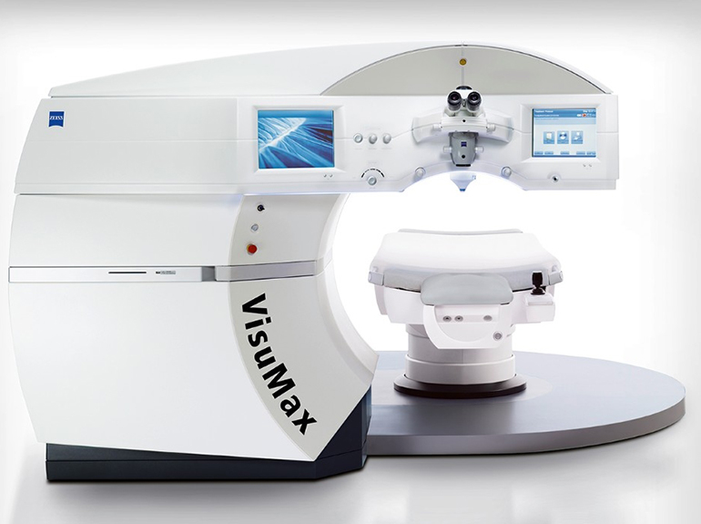
With ReLEx® from SMILE, a lenticule and a small incision of < 6 mm is created inside the intact cornea, enabling an 80% smaller side-cut and 30% smaller cap cut, as compared to a LASIK flap. With ZEISS ReLEx SMILE, the entire correction is performed as a single-step: one laser, one treatment plan and one laser process.
The VisuMax is the first laser system to perform the minimally invasive, flapless SMILE procedure. With ReLEx SMILE from ZEISS, a refractive lenticule, as well as the incision through which it is extracted, are created in a single step – without ablation or flap.
WAVELIGHT EX500 EXCIMER LASER
.jpg)
The ultraviolet light from an excimer laser is well absorbed by biological matter and organic compounds. Rather than burning or cutting material, the excimer laser adds enough energy to disrupt the molecular bonds of the surface tissue, which effectively disintegrates into the air in a tightly controlled manner through ablation rather than burning.Thus excimer lasers have the useful property that they can remove exceptionally fine layers of surface material with almost no heating or change to the remainder of the material which is left intact. These properties make excimer lasers well suited to precision micromachining organic material (including certain polymers and plastics), or delicate surgeries such as eye surgery LASIK.
WHITESTAR SIGNATURE PRO

It is a method of cataract removal by use of ultrasonic emulsification with aspiration of the cataractous lens through a small incision. The WHITESTAR SIGNATURE PRO System represents the latest generation of AMO phacoemulsification technology. Designed and manufactured into every WHITESTAR SIGNATURE PRO System are safety, ease-of-use, and reliability. The WHITESTAR SIGNATURE PRO System meets applicable United States and International safety requirements for this type of device.The WHITESTAR SIGNATURE PRO System contains a number of features based on extensive research and clinical trials with highly trained and noted ophthalmologists with experience as phacoemulsification surgeons.
CENTURION VISION SYSTEM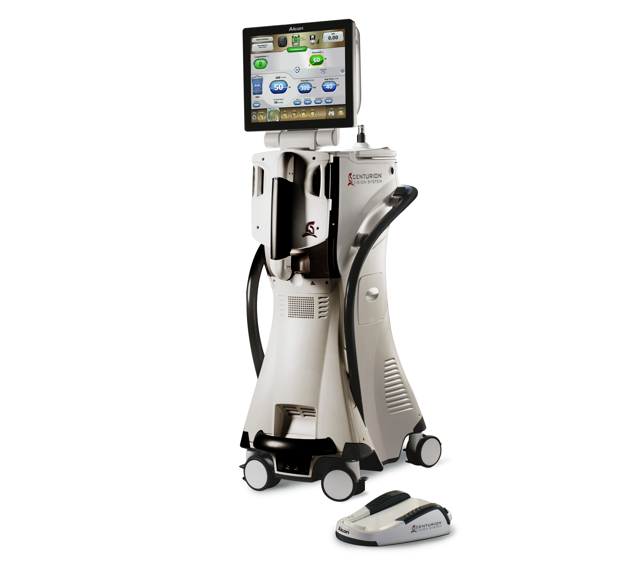
CENTURION® Vision System a new phacoemulsification (phaco) technology platform designed to provide surgeons improved capabilities in cataract removal.The CENTURION Vision System sets a new standard of performance in cataract surgery by combining multiple intelligent phaco technologies, and other key features.
LUMERA SURGICAL MICROSCOPE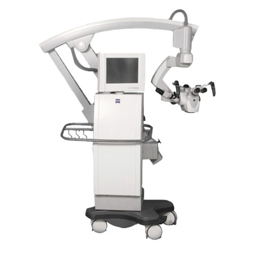
The OPMI Lumera ophthalmic surgical microscope is high end in everything but price. Equipped with SCI(TM) (Stereo Coaxial Illumination) for a brilliant red-reflex critical for anterior segment work, legendary Carl Zeiss apochromatic optics for unparalleled visual clarity and ergonomic considerations like a touch screen graphical user interface that allows intuitive control of all system functions, including the integrated video camera, the OPMI Lumera is the perfect scope for surgery centers and office alike.
B SCAN (BRIGHTNESS SCAN)
B-scan, or brightness scan, is another method used for ocular assessment via ultrasound. It can be performed directly on the anesthetized eye.
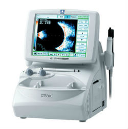
In cases of trauma or in children, B-scan can be performed over the eyelid with coupling jelly. Measurements derived from B-scan include visualization of the lesion, including anatomic location, shape, borders, and size. It can be used for a detection of a wide-range of pathological structures, including retinal or choroidal detachment, foreign bodies, calcium, and tumors.
Pharmacy and Optical Store


